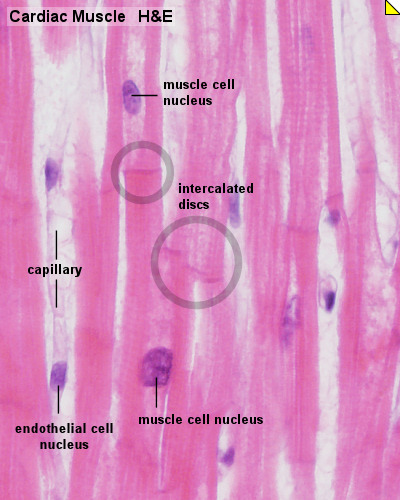
Cardiac Muscle Histology Slide Labeled
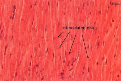
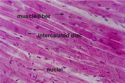
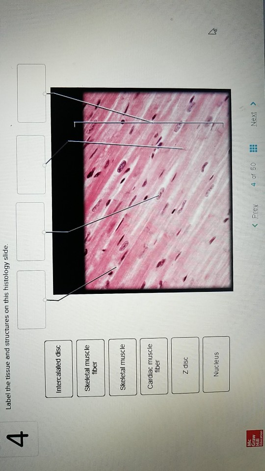
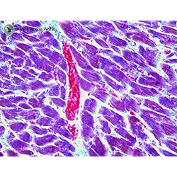
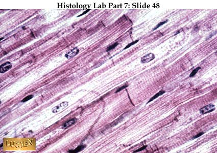
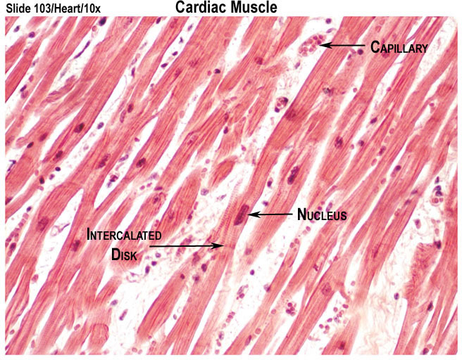
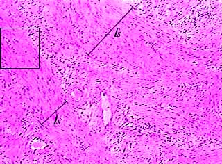
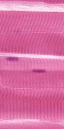
Cardiac muscle 40x the individual cardiac muscle cells are arranged in bundles that form a spiral pattern in the wall of the heart.
:background_color(FFFFFF):format(jpeg)/images/library/3490/5heSmCkewUh8Qv1PWQNM2Q_Z_Lines.png)
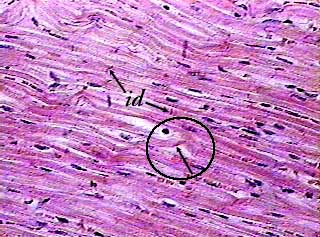
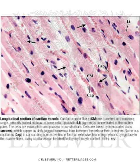
Cardiac muscle histology slide labeled. Histology of cardiac muscle 1. 305 heart ventricle he webscope note. The cells are often branched and are tightly connected by specialised junctions. Cardiac muscle tissue also known as myocardium is a structurally and functionally unique subtype of muscle tissue located in the heart that actually has characteristics from both skeletal and muscle tissuesit is capable of strong continuous and rhythmic contractions that are automatically generated.
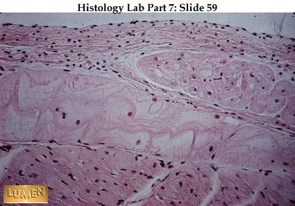
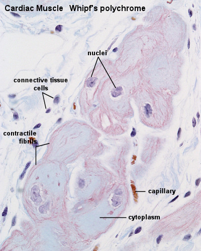
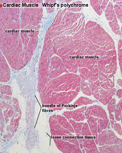
This slide not in glass slide collection cardiac muscle will be studied in the wall of the ventricle of the heart. The contractility can be altered by the autonomic nervous system and hormones. Histology of cardiac muscle 1 by. Purkinje fibre sheep whipfs polychrome cardiac muscle cells in this preparation have a red violet appearance.
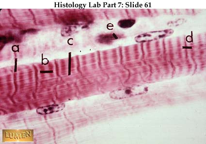
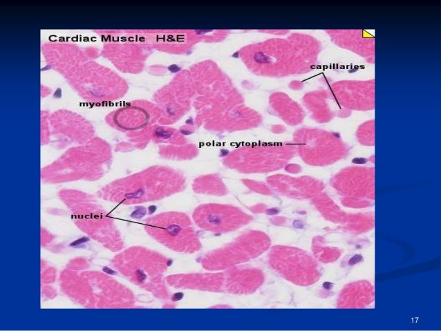
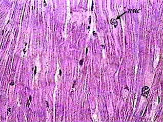
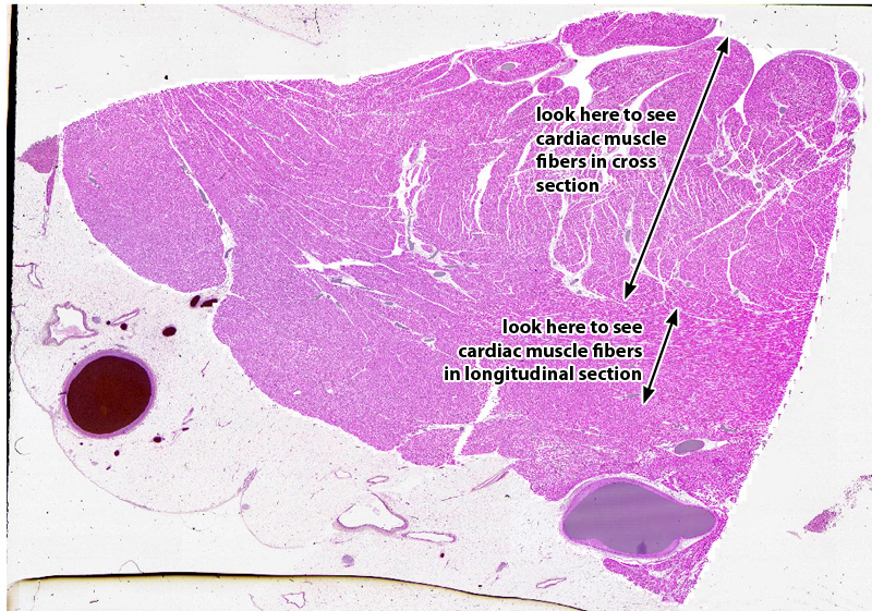
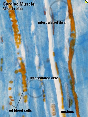
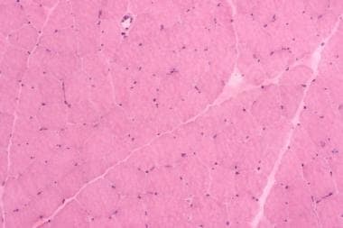
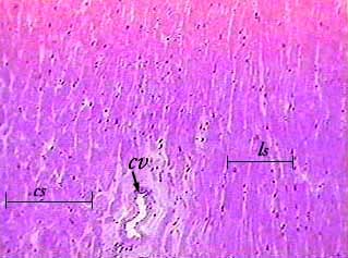
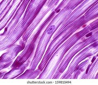
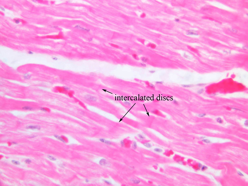
When cardiac muscle fibers contract not only do the muscle fibers shorten but the spiral bundles twist to compress the contents of the heart chambers. On any slide of cardiac muscle you will see cells that have been sectioned in every possible direction from transverse to oblique to longitudinal. Much of the connective tissue looks light blue striations of cardiac muscle cells are visible. Cardiac muscle cells or cardiomyocytes contain the same contractile filaments as in skeletal muscle.
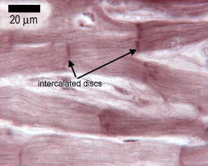
Blood is an important medium that not only carries nutrients and oxygen throughout the body but it also collects waste products and returns them to the liver and kidney for further processing and excretion. Cardiac muscle cells excitation is mediated by rythmically active modified. Cardiac muscle is striated like skeletal muscle as the actin and myosin are arranged in sarcomeres just as in skeletal muscle. However cardiac muscle is involuntary.
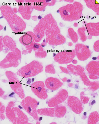
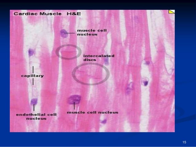
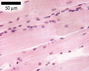
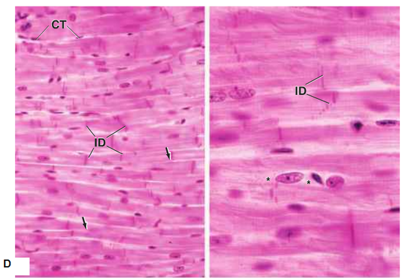
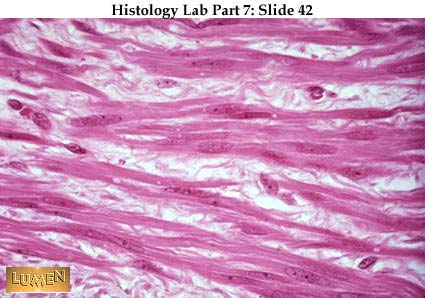
Cardiac muscle cells usually have a single central nucleus. In comparison with skeletal muscle note the following differences. Introduction cardiac muscle the myocardium consists of cross striated muscle cells cardiomyocytes with one centrally placed nucleus. Cardiac muscle is striated involuntary muscle found in the heart wall.
Cardiac muscle cells have rounded cross sections less than 25 um in diameter with a centrally located nucleus. Suitable slides sections of cardiac muscle interventricular septum whipfs polychrome iron haematoxylin he. Nuclei are oval rather pale and located centrally in the muscle cell which is 10 15 um wide. The heart is able to achieve this autonomy based on its histological make up.


:background_color(FFFFFF):format(jpeg)/images/article/en/cardiac-tissue/Dlly3NxuFvJnfdwmsCvVrQ_LNOsY5VQ7ADcaM1g9m5g_Cardiac_Muscle.png)

:background_color(FFFFFF):format(jpeg)/images/library/7173/Skeletal_muscle_01.png)
















_040_02.jpg)


:background_color(FFFFFF):format(jpeg)/images/library/3159/W8vCwHUn3D60Kb5xvvzvXQ_Intercalated_discs.png)











:background_color(FFFFFF):format(jpeg)/images/article/en/histology-of-skeletal-muscle/CCA8JH8f29ZhIfXBU6FQ_09gjtxSIpcGC6ni5oqNdg_Striations.png)

